Slider Background

Main Text


Slogan Section

Service Section


Why choose us?

We are the best medical, ethical and legal choice for professionals who want to opt for technology.


Our lab

In Hearten Biotech, our laboratory has been developed according to the highest standards of biosecurity.


Products

We develop products for the treatment of various pathologies that can affect an dogs, cats and horses.

Tissue engineering and cell therapies are powerful tools for the treatment of acute and chronic diseases.
More information

News

All

News

Events

domingo, 21 de agosto de 2016

Electricity and Stem Cells: Old Signal for New Therapies


August 2016
Jorge Genovese, MD PhD

Electrical Stimulation Induces Myogenic Differentiation of Human Dermal Fibroblasts.
From Dr. Genovese's lab: After 12 hours of in vitro 12V electrical stimulation (image on right), dermal fibroblasts express key muscle proteins: cardiac troponin (green), MyoD (green spots on blue nuclei), and connexin-43 (brown).
Stem cells are very sensitive sensors, able to detect a myriad of minimal changes in their environment. In vitro and in vivo, stem cells react to different stimuli by migrating, dividing, dying, changing their secretory profile and/or modifying their own shape.
All these actions are the detectable expression of cell’s ability to quickly and specifically modify their gene expression. This dramatic modulation of genetic information and its translation has as its main objective to restore normal function  at the site of an injury.
Electrical stimulation is a well-known trophic factor for different tissues. Endogenous bioelectric currents are involved in normal tissue function and repair.  Specific ion channels and pumps within cell membranes generate bioelectric signals. Its action is evident, but not limited to, the nervous system and muscular tissue. Although the mechanisms underlying the effects of electrical stimuli are not totally understood, it is clear that they affect cellular proliferation, hypertrophy and apoptosis [1]. 
Electrical stimulation or “electrotherapy” is widely applied in human medicine, especially in neuromuscular disorders. Electricity is used in rehabilitative medicine due to its ability to restore muscle function through induction of new blood vessel formation  - angiogenesis - and the promotion of cell proliferation [2].  In a similar manner, electrical treatment improves wound healing and osteogenesis. All previous data indicate a clear involvement of stem cells in this regenerative process, and an action of electrical stimulation on stem cells. 
Cell movement and cell positioning are important components of regeneration and the right bioelectric signals can get regenerative repair cells to where they need to be.  Bioelectric signals can turn up or turn off proliferation.  They can cause new blood vessels to grow or can suddenly halt blood supply, as may be needed in the case of starving cancer tumors. Certain bioelectric signals can even affect cell elimination through programmed cell death. Experiments have proven the ability of bioelectric stimulation to induce or augment regeneration, which is our area of greatest research interest.
Electrical stimulation induces dramatic changes in stem cell activity toward a clear regenerative phenotype.  Exogenous electrical currents activate and mobilize neural stem cells in vitro and in vivo. Invasive and non-invasive electrical stimulation of the central nervous system have showed, both in animal models and in patients, enhancement of neurogenesis and cellular plasticity [3].  
Depending on the intensity of the electrical stimulation they receive, mesenchymal stem cells proliferate and modify their phenotype. Short time treatments let stem cells pre-differentiate into cardiac cells [4] . Preconditioning treatment with electricity promotes stem cell survival post-transplantation, enhancing their beneficial effects.  Electrical stimulation induces the production of cardiac proteins that facilitate cell engraftment and survival. This action is not limited to the heart or to mesenchymal stem cells. Induced pluripotent stem cells respond to electrical stimulation in a similar manner, and fibroblasts, the main cell type in connective tissue, show cardiac differentiation as well [1].
Electrical stimulation acts directly and indirectly on stem cells. In a rat model, when the stimuli were delivered across a post-infarct scar, an enhanced presence of hematopoietic stem cells in the lesion was detected. These cells appear to be associated with an increase of local vascular endothelial growth factor levels and new vascular structures [5]. Today, we accept that cells associated with the vessel wall – pericytes - are the most relevant mesenchymal stem cell population. In this way, electrical stimulation thus not only induces proliferation and differentiation but the recruitment of different stem cell types in the area to repair the lesion.
Electro stimulation of scaffolds and/or cell-scaffold constructs opens a new area in tissue engineering developments. We are optimistic that the combination of physical and biochemical signals with stem and non-stem cells can accelerate the generation of artificial tissues, specifically for cardiac and skeletal muscle.
“Electroceuticals” is a word that encompasses all medical devices which employ electrical stimulation to affect and modify functions of the body. In near future medicine, miniaturized electroceutical devices will open a new perspective. Many of these devices act on stem cells through the mechanisms above mentioned. Applied on body’s surface or implanted in target organs, they will deliver specific electrical stimuli to modulate stem cells number, differentiation grade, biological activity and even proliferation or survival. 
Electricity and electroceutical devices appear to be a selective non-drug approach to resolve a huge amount of physical conditions. The regulation of hair follicle activity, the induction of muscle proliferation in a weak aneurysm wall, the differentiation of neural progenitors after stroke, or the induction of differentiation of cancer stem cells, are just some applications of this impressive therapeutic tool.
By learning to modulate the bioelectrical signals that control regeneration promoting protein expression, stem cell recruitment, cell proliferation and differentiation, we may gain an incredible new set of tools to repair failing organs to not only extend life but restore a high quality of life with full functioning regenerated organs. 
Jorge Genovese, M.D., Ph.D., is the President of Hearten Biotech  and Director of Electrical Stimulation Regeneration Research at BioLeonhardt Inc. Dr. Genovese obtained his M.D. and Ph.D. from Buenos Aires University. He has been a faculty member of the McGowan Institute for Regenerative Medicine at the University of Pittsburgh, where he was the Principal Investigator at the Center for Cardiac Cell Therapy and the Cardiac and Molecular Biology Laboratory. He was Director of the Cardiac Regenerative Medicine Laboratory at the Cardiac Surgery Division, University of Utah. Dr. Genovese was also Invited Cardiac Surgery Professor at Campus Biomedico University in Rome. Dr. Genovese has been very active in the Tissue Engineering International & Regenerative Medicine Society (TERMIS), chairing numerous committees and serving a term as Vice President. He is Editor of the Journal of Stem Cells, Associated Editor of the Frontiers in Stem Cells Journal, member of the Editorial Committee of the World Journal of Stem Cells, and member of the North American Veterinary Regenerative Medicine Association. Dr. Genovese is a pioneer of Tissue Engineering in Latin America, being the first in the region to generate keratinocytes cultures in 1985, an organotípico dermoepidermic device in 1998, and a genetic modified dermo-epidermic device in 2002, among many other tissues.

References

  1. J.A. Genovese, C. Spadaccio, J. Langer, J. Habe, J. Jackson, and A.N. Patel, Electrostimulation induces cardiomyocyte predifferentiation of fibroblasts. Biochemical and Biophysical Research Communications370 (2008) 450-5. doi:10.1016/j.bbrc.2008.03.115
  2. U. Carraro, K. Rossini, W. Mayr, and H. Kern, Muscle fiber regeneration in human permanent lower motoneuron denervation: relevance to safety and effectiveness of FES-training, which induces muscle recovery in SCI subjects. Artificial Organs 29 (2005) 187-91. doi:10.1111/j.1525-1594.2005.29032.x
  3. Y. Huang, Y. Li, J. Chen, H. Zhou, and S. Tan, Electrical Stimulation Elicits Neural Stem Cells Activation: New Perspectives in CNS Repair. Frontiers in Human Neuroscience 9 (2015) 586. doi: 10.3389/fnhum.2015.00586
  4. J.A. Genovese, C. Spadaccio, E. Chachques, O. Schussler, A. Carpentier, J.C. Chachques, and A.N. Patel, Cardiac pre-differentiation of human mesenchymal stem cells by electrostimulation. Frontiers Bioscience (Landmark Ed) 14 (2009) 2996-3002. [PDF]
  5. C. Spadaccio, A. Rainer, F. De Marco, M. Lusini, P. Gallo, P. Sedati, A.O. Muda, S. De Porcellinis, C. Gregorj, G. Avvisati, M. Trombetta, M. Chello, E. Covino, D.A. Bull, A.N. Patel, and J.A. Genovese, In situ electrostimulation drives a regenerative shift in the zone of infarcted myocardium. Cell Transplant22 (2013) 493-503. http://dx.doi.org/10.3727/096368912X652977
Leer más

lunes, 25 de julio de 2016

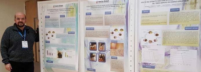
Hearten Biotech has been in SOLCEMA's IV Congress: " Células madre: Nueva Era de la Medicina", held in Viña del Mar, Chile from the 20th till the 24th of July. The main purpose was to present three different cases in canine treated with Stem Cells by Hearten Biotech. The diagnosis were Early Severe Vasculitus, Chronic Pancrestitis and Epilepsy.
We want to thank for giving us the opportunity to be there and thank Dr. Jorge Genovese, our CEO, who represented Hearten Biotech at the Congress.
Dr. Genovese and our Stem Cells treatments. 


Leer más

viernes, 22 de enero de 2016

La investigación comenzó con sólo una ínfima ‪plaqueta‬ de la sangre, pero pronto descubrieron grandes oportunidades para el crecimiento y la amplitud de su investigación.

Según Bulla, el cáncer necesita la creación de nuevos vasos sanguíneos, llamados angiogénesis, para sobrevivir y crecer, y los tumores son capaces de crear nuevos vasos sanguíneos como vías para transportarse y propagarse. Ellos también están estudiando la manera en que las plaquetas interactúan con las células tumorales en su intento de extenderse a la zona que rodea el tumor o metástasis a sitios distantes en el cuerpo.

Lunsford, profesor asociado en el departamento de ciencias de la clínica, dijo: "Sabemos que las ‪#‎células‬ tumorales que se diseminan por metástasis, necesitan a las plaquetas, pero aún no se sabe qué hacen las plaquetas por el tumor (metástasis) de migración. Esta es una de las preguntas que esperamos ayudar a responder”.

"Si los tratamientos son exitosos y el cáncer entra en remisión, se supervisaría el paciente para una recaída de la enfermedad observando sus plaquetas", dijo Lunsford. "Este tipo de monitoreo sería menos invasivo que la toma de biopsias y también podría ser un indicador temprano de que el cáncer está regresando."

Según Lunsford, las plaquetas también contienen información acerca de los tumores y las células metastásicas del cáncer, y el equipo espera que mirando las proteínas específicas expresadas en las plaquetas (a partir de una simple muestra de sangre), pueden identificar cáncer con antelación. Aún más importante, quieren identificar cuando los tumores están a punto de hacer metástasis.

Sobre la base de lo que han aprendido en Harvard y su propio trabajo en la MSU, los investigadores dijeron que sienten que hay una clara relación entre la enfermedad en los animales y los seres humanos. Sus esfuerzos también son parte de la Iniciativa de Salud Uno, un programa mundial actual para expandir colaboraciones interdisciplinarias y las comunicaciones en todos los aspectos de la atención de salud para los seres humanos, los animales y el medio ambiente.

Fuente: http://www.sciencedaily.com/releases/2014/10/141030213702.htm
Leer más
M.V. Luciana Monteiro Mazzola
Entrevistamos a la Médica Veterinaria Luciana Monteiro Mazzola para que nos explique qué es la Medicina Regenerativa, y como se aplica al Universo Veterinario.


Durante los últimos años los avances en el ámbito de la biología molecular y celular, han ganado cada vez más apoyo como  método  eficaz para restaurar o mejorar la función de órganos y tejidos lesionados por ciertas enfermedades o traumatismos. Así es como la medicina tradicional está cambiando su paradigma, vinculado estrechamente a los nuevos conocimientos sobre las células madre y poniendo a la Medicina Regenerativa en un primer plano.

Este promisorio campo se extiende a todas las ramas de la medicina y toma un particular protagonismo en la Veterinaria. Para entender un poco más de cómo puede ayudar y cambiar la vida cotidiana de nuestros animales hablamos con la Médica Veterinaria Luciana Monteiro Mazzola.


Células Madre, Terapia Celular, Ingeniería de Tejidos…hace tiempo que estos temas resuenan en el mundo, pero su aplicación práctica en veterinaria es relativamente nueva.  ¿En qué punto del proceso de estandarización de los tratamientos nos encontramos?

La aplicación de la medicina regenerativa no es nueva, ya hace varios años que se está utilizando. En Estados Unidos es de uso frecuente  y actualmente hay cada vez más países que están incorporando estas exitosas herramientas terapéuticas.

El objetivo concreto de la medicina regenerativa es reemplazar o regenerar las células, tejidos u órganos para conseguir restaurar o restablecer su función normal. Esto se logra mediante la estimulación de los propios mecanismos del cuerpo para reparar esos tejidos dañados,  regenerando parte de los mismos en el animal, o reemplazándolos completamente por tejidos u órganos creados in vitro.

En cuanto a tu pregunta de  en qué punto nos encontramos, considero que se han logrado grandes avances en el campo de la medicina regenerativa pero aún quedan muchas cosas más por hacer.

¿Qué hay de cierto en que una célula madre podrá corregir cualquier defecto del órgano en el que se aplique?

Si bien existen diversas indicaciones terapéuticas,  no creo que puedan corregir cualquier defecto en el órgano a tratar.  Sus efectos dependerán del momento de la aplicación, órgano afectado, tipo y  origen de la lesión.

Y en base a los casos actuales que se le presentan, ¿Cuáles son los problemas por los que más consultan los veterinarios?

Actualmente los problemas ortopédicos tienen una alta incidencia en la clínica y hoy existen diversos trabajos que prueban la eficiencia del uso de estos productos en su tratamiento.

La artrosis, por ejemplo, es una patología en la que ya se está aplicando la terapia con células madre de forma exitosa, tanto en humanos como en veterinaria.

Usted estuvo trabajando como veterinaria en el exterior, en su experiencia, ¿cuál es la situación actual de Argentina con respecto a otros países?

En la Argentina las terapias celulares son relativamente nuevas y aun no están incorporadas a la práctica clínica de la mayoría de mis colegas. En Hearten creemos que en los próximos años esto cambiará gracias a los grandes avances que se están obteniendo en los países centrales.

Actualmente usted se encuentra trabajando con un referente en el tema, el Dr. Jorge Genovese. ¿Qué  le despertó tanto interés y entusiasmo en la terapia celular?

Cuando surgió la posibilidad de incorporarme al equipo de Jorge,  ni lo dude. A parte de ser un pionero en el área de la Medicina Regenerativa,  es un gran maestro para todos los que estamos en la compañía.

Lo que despertó mi entusiasmo por incorporar estas herramientas terapéuticas a la clínica veterinaria fue la gran cantidad de trabajos publicados acerca del uso del PRP, fracción vascular de la grasa y células madres mesenquimales. Espero que podamos tener cada vez más veterinarios que utilicen y comprueben  la eficiencia de nuestros productos.

¿Y  por qué cree que hay tanta desconfianza o prejuicio frente al tema?

Posiblemente por desconocimiento y porque aun no está afianzado el uso de estos productos en Argentina.
Leer más

Nos complace anunciarles que el día de hoy el Dr. Jorge Genovese impartirá una charla sobre La medicina veterinaria regenerativa a las 10 pm horario de Argentina.  A continuación el link por el cual podrá ver esta interesante charla.


http://veterinariosenweb.com/campus/vivo/2015/programa_vetes_en_vivo.html


Leer más

Agradecemos al Dr. Juan Pablo Quiroga, veterinario de La Dolfina, por contar con nosotros para el cuidado de sus pacientes y valoramos su experticidad y profesionalismo.


Gracias
Leer más

jueves, 26 de marzo de 2015

El Instituto Nacional de Enfermedades Virales Humanas “Dr. Julio I. Maiztegui” dictará su renombrado curso “EL CULTIVO CELULAR COMO HERRAMIENTA EN MEDICINA HUMANA Y VETERINARIA” los días 13, 14, 15 y 16 de Abril de 2015 en la ciudad de Pergamino (Bs. As).

El curso se orienta a capacitar a los participantes en todos los aspectos básicos del manejo del cultivo celular, así como en la organización de los componentes del entorno que influyen en la calidad, trazabilidad y sustentabilidad de los productos obtenidos.

El curso contará con la disertación del Dr. Jorge Genovese, quien hablará sobre la utilización de Células Madre en Terapias y Medicina Regenerativa.

Para acceder al programa del curso ingrese aquí.

Para mayor información puede escribir a cursosinevh@gmail.com.
Leer más